Opening comparison and immediate clinical trigger
Practices choose a scanner not because it’s new, but because it solves a specific problem: speed, fit, or predictable occlusion. For instance, crown-and-bridge workflows demand reliable bite data; a stable jaw relation record early in the scan cuts remakes. I’ll compare the practical trade-offs so you can match a device to daily needs rather than marketing claims.

Core technologies and what they mean for outcomes
Most scanners use structured light or confocal imaging. Structured light is fast and forgiving on texture; confocal systems often give higher point density on enamel. Consider scanning strategy: some excel at single-unit detail, others handle full-arch stitching better. When you compare devices, look at real-world scan speed, how the software stitches images, and whether the system compensates for patient movement.

Clinical applications with direct comparisons
Restorative: For single crowns, a scanner with high resolution and reliable margin capture matters more than raw speed. Orthodontics: you prioritise full-arch repeatability and integration with aligner labs. Implants: accurate implant position and soft-tissue capture reduce abutment adjustments. Prosthetics and full-arch: pick a system proven for long-span stitching and occlusal relationships. Match the scanner’s strengths to the task instead of forcing one device to cover all roles.
Workflow integration: open vs closed, CAD/CAM and lab handoffs
Open systems export standard file types and make lab collaboration smoother. Closed systems may offer tighter in-house chains but can lock you into proprietary workflows. Compare how easily the scanner exports STL/PLY/OBJ, how APIs or plugins connect to your CAD software, and whether the vendor updates drivers regularly. A cleaner file handoff reduces adjustments and clarifies responsibility between clinic and lab.
Accuracy, repeatability and common scanning mistakes
Accuracy is task-specific: implant position for a surgical guide demands different tolerances than a temporary crown. Repeatability matters more than a single “best” measurement. Watch for common mistakes—insufficient soft-tissue retraction, reflective restorations, incomplete margins, and poor bite capture. Train staff on scan path, use anti-reflective sprays only when indicated, and verify occlusion in segments rather than relying on a single full-arch capture.
Patient experience, ergonomics and chairside practicalities
Probe size, cable length, and heat affect patient comfort and operator fatigue. Short learning curves speed adoption: a system that’s intuitive for hygienists lets you delegate routine impressions. Also, consider infection control: detachable tips that tolerate autoclaving make workflows simpler. Compare user interfaces; a slow touchscreen or laggy software adds minutes to each appointment.
Cost, service and measurable return
Cost isn’t just initial purchase. Factor in service contracts, software updates, training, and consumables. Estimate time savings: if a scanner shortens impression and lab turnaround by predictable minutes per case, you’ll see capacity gains. Insist on vendor transparency about upgrade costs. Compare warranties and local technical support responsiveness before committing.
Alternatives and when to keep analog or add desktop scanning
Some clinics retain conventional impressions for specific cases: extreme subgingival margins or severe trismus remain challenging for intraoral devices. Desktop scanners still excel at die accuracy for complex lab work. Choose a hybrid approach if your caseload is mixed—use intraoral scanning for routine cases and send impressions to the lab when digital capture risks compromise.
Industry signals and a real-world anchor
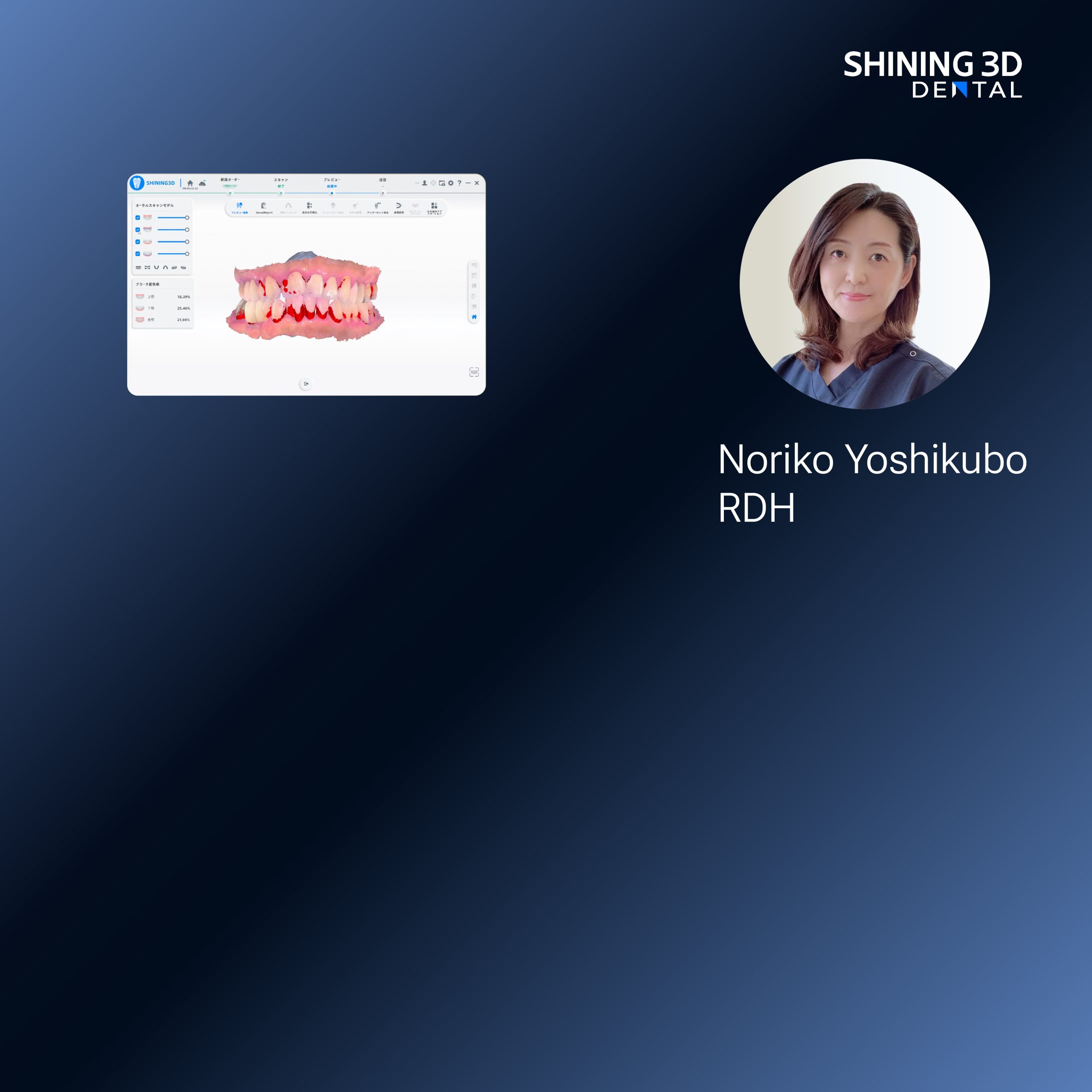
At the IDS in Cologne, vendors demonstrated tighter integrations between scanners and restorative workflows, including demonstrations of a future digital dental solution that focused on occlusal and jaw relation consistency. These presentations highlighted practical interoperability more than raw specs: how a system plugs into daily practice determines its value.
Final synthesis and practical takeaway
Compare devices by task: choose detail for single restorations, stitching reliability for full-arch work, and open export for lab collaboration. Watch training, support, and real-case ergonomics more than benchmark numbers. When you match scanner strengths to clinical needs, you reduce remakes and save clinical time—outcomes that matter to patients and teams alike, as demonstrated by modern solutions from SHINING 3D DENTAL.